M1: Identify the functions.
In order for neurons to become active, they must receive action potentials or other stimuli. Dendrites are the structures on the neuron that receive electrical messages. These messages come in two basic forms: excitatory and inhibitory.
The function of the axon is to transmit information to different neurons, muscles and glands.
Axomembrane is the collection of dendrites and axons.

M2: Distinguish among sensory, motor and interneurons.
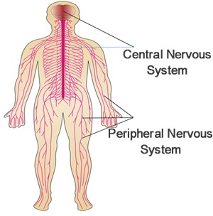
Sensory neuron: type of neuron which takes a message from a sense organ to the CNS
Motor neuron: type of neuron which takes a message away from the CNS to an effector (muscle or gland), it has short dendrites and a long axon
Interneuron: type of neuron found completely within CNS which conveys messages between parts of the system; has short dendrites and long/short axon


M3: Explain transmission of a nerve impulse through a neuron, using the following terms:
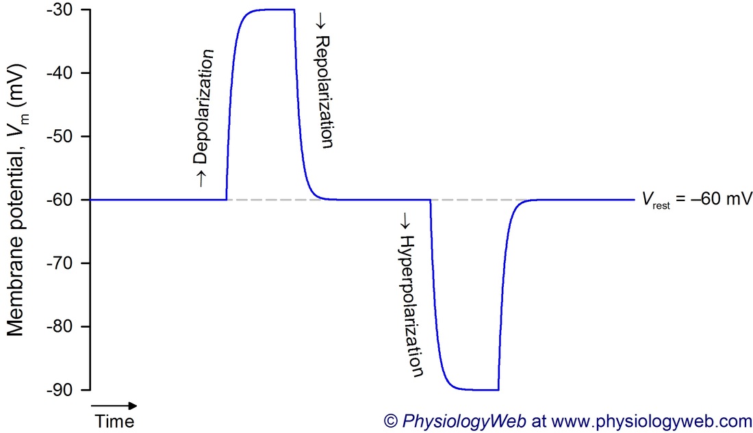
When a neuron is not sending a signal, it is "at rest." When a neuron is at rest, the inside of the neuron is negative relative to the outside. Although the concentrations of the different ions attempt to balance out on both sides of the membrane, they cannot because the cell membrane allows only some ions to pass through channels (ion channels). At rest, potassium ions (K+) can cross through the membrane easily. Also at rest, chloride ions (Cl-)and sodium ions (Na+) have a more difficult time crossing.
After a cell has been depolarized, it undergoes one final change in internal charge. Following depolarization, the voltage gated sodium ion channels that had been open while the cell was undergoing depolarization close again. The increased positive charge within the cell now causes the potassium channels to open.
Sodium-potassium pump: a protein carrier which pumps Na+ out and K+across the axon membrane. It is always working.
Recovery period: also known as a refractory period whereby a fiber cannot conduct an impulse; occurs right after an impulse has been conducted; insures one-way direction of impulse.
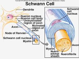
M4: Relate the structure of a myelinated nerve fiber to the speed of the impulse conduction.
Schwann cells are known for their roles in supporting nerve regeneration. Nerves in the PNS consist of many axons myelinated by Schwann cells. If damage occurs to a nerve, the Schwann cells will aid in digestion of its axons. Schwann cells (neurolemmocytes) are neuroglial cells which encircle a fiber, leaving gaps called the nodes of Ranvier (neurofibril nodes). Schwann cells wrap themselves around the axon many times, and in this way lay down several layers of plasma membrane containing myelin, which forms a myelin sheath.
Schwann cells are known for their roles in supporting nerve regeneration. Nerves in the PNS consist of many axons myelinated by Schwann cells. If damage occurs to a nerve, the Schwann cells will aid in digestion of its axons. Schwann cells (neurolemmocytes) are neuroglial cells which encircle a fiber, leaving gaps called the nodes of Ranvier (neurofibril nodes). Schwann cells wrap themselves around the axon many times, and in this way lay down several layers of plasma membrane containing myelin, which forms a myelin sheath.
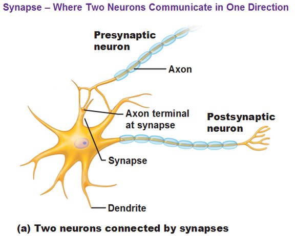
M5: Identify the major components of a synapse
Axon bulb is the small swelling on an axon branch, lying very close to the dendrite/cell body of another nuron. Presynaptic membrane is the membrane of the first neuron (the axon). Postsynaptic membrane is the membrane of the next neuron
Presynaptic relating to or denoting a nerve cell that releases a transmitter substance into a synapse during transmission of an impulse.
Postsynaptic membrane is the membrane of the next neuron (the dendrite).
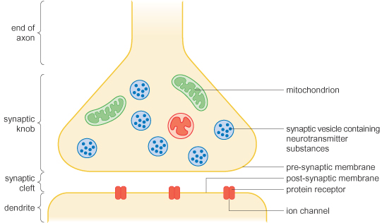
Synaptic cleft is the small gap between the presynaptic and postsynaptic membrane.
Neurotransmitters are chemicals stored at then ends of axons in vesicles that is responsible for transmission across a synapse, by causing excitation (causes Na+ channels to open; neuron transmits a nerve impulse)
Axon bulb is the small swelling on an axon branch, lying very close to the dendrite/cell body of another nuron. Presynaptic membrane is the membrane of the first neuron (the axon). Postsynaptic membrane is the membrane of the next neuron
Presynaptic relating to or denoting a nerve cell that releases a transmitter substance into a synapse during transmission of an impulse.
Postsynaptic membrane is the membrane of the next neuron (the dendrite).
Synaptic cleft is the small gap between the presynaptic and postsynaptic membrane.
Neurotransmitters are chemicals stored at then ends of axons in vesicles that is responsible for transmission across a synapse, by causing excitation (causes Na+ channels to open; neuron transmits a nerve impulse)
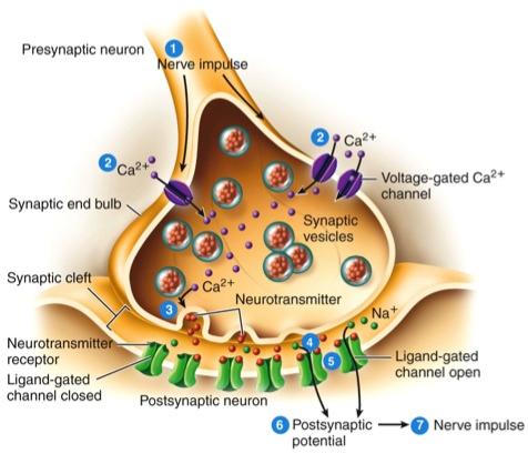
M6: Explain the processes by which impulses travel across a synapse
When a nerve impulse reaches the synapse at the end of a neuron, it cannot pass directly to the next one. Instead, it triggers the neuron to release a chemical neurotransmitter. The neurotransmitter drifts across the gap between the two neurons.
When a nerve impulse reaches the end of an axon, the axon releases chemicals called neurotransmitters . Neurotransmitters travel across the synapse between the axon and the dendrite of the next neuron. Neurotransmitters bind to the membrane of the dendrite.
M7: Demonstrate knowledge of how transmitters are broken down in the synaptic cleft
The postsynaptic membrane contains enzymes that rapidly inactivate the neurotransmitter. For example, acetycholinesterase (AChE) breaks down acetylcholine. Sometimes, the synaptic ending rapidly reabsorbs the neurotransmitter, for repackaging in a synaptic vesicle or for chemical breakdown.
The postsynaptic membrane contains enzymes that rapidly inactivate the neurotransmitter. For example, acetycholinesterase (AChE) breaks down acetylcholine. Sometimes, the synaptic ending rapidly reabsorbs the neurotransmitter, for repackaging in a synaptic vesicle or for chemical breakdown.
M8: Relate the structure of a reflex arc to how it functions.
Reflex Arc: the nerve pathway involved in a reflex action including at its simplest a sensory nerve and a motor nerve with a synapse between.
When a reflex arc consists of only two neurons in an animal (one sensory neuron, and one motor neuron), it is defined as monosynaptic. Monosynaptic refers to the presence of a single chemical synapse. In the case of peripheral muscle reflexes (patellar reflex, achilles reflex), brief stimulation to the muscle spindle results in contraction of the antagonist or effector muscle.
By contrast, in polysynaptic reflex pathways, one or more interneurons connect afferent (sensory) and efferent (motor) signals. All but the most simple reflexes are polysynaptic, allowing processing or inhibition of polysynaptic reflexes within the spinal cord.