The main types of blood vessels are:
- Arteries: any of the muscular-walled tubes forming part of the circulation system by which blood (mainly that which has been oxygenated) is conveyed from the heart to all parts of the body.
- Arterioles: is a small diameter blood vessel in the microcirculation that extends and branches out from an artery and leads to capillaries. Arterioles have muscular walls (usually only one to two layers of smooth muscle) and are the primary site of vascular resistance.
- Capillaries: any of the fine branching blood vessels that form a network between the arterioles and venules.
- Venules: a very small vein, especially one collecting blood from the capillaries.
- Veins: any of the tubes forming part of the blood circulation system of the body, carrying in most cases oxygen-depleted blood toward the heart.

J2: List the following functions.
There are two subclavian arteries that supply our arms with blood. The subclavian arteries branch to the vertebral arteries. These carry oxygenated blood up to the brain from the base of the neck. The right subclavian artery is located below the clavicle.
There are two jugular veins on each side of the neck, known as the external and internal jugulars. The external one lies close to the surface and carries blood from the outside parts of the head and neck to the heart.
The carotid arteries are major blood vessels in the neck that supply blood to the brain, neck, and face. There are two carotid arteries, one on the right and one on the left.
The superior mesenteric artery is a major blood vessel in the digestive system. This artery branches off the abdominal aorta and supplies oxygenated blood to the pancreas and the lower parts of the intestine.
The pulmonary veins are large blood vessels that receive oxygenated blood from the lungs and drain into the left atrium of the heart.
The hepatic veins carry oxygen-depleted blood from the liver to the inferior vena cava. They also transport blood that has been drained from the colon, pancreas, small intestine, and the stomach, and cleaned by the liver.
The portal vein or hepatic portal vein is a blood vessel that conducts blood from the gastrointestinal tract and spleen to the liver.
The internal iliac artery, also called the hypogastric artery, is the dominant artery in the pelvic area. It is usually shorter in length than the external iliac artery.
Coronary arteries supply blood to the heart muscle. Like all other tissues in the body, the heart muscle needs oxygen-rich blood to function, and oxygen-depleted blood must be carried away.
The aorta is the largest artery in the body. The aorta begins at the top of the left ventricle, the heart's muscular pumping chamber. The heart pumps blood from the left ventricle into the aorta through the aortic valve.
J3: Demonstrate safe dissection techniques.
https://www.youtube.com/watch?v=49LjT7upENM
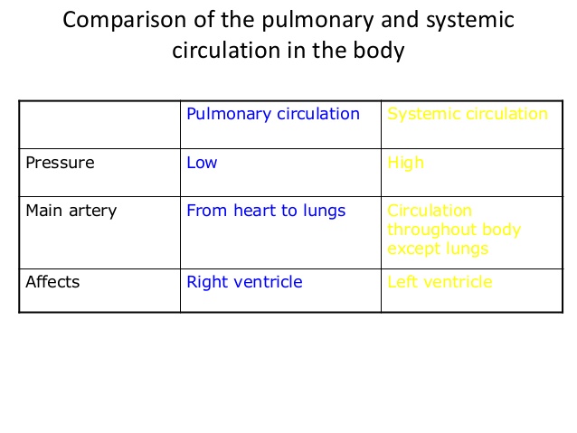
J4: Difference between Pulmonary and Systemic circulation.
Systemic circulation is the movement of blood from the heart through the body to provide oxygen and nutrients, and bringing deoxygenated blood back to the heart. Oxygen-rich blood from the lungs leaves the pulmonary circulation when it enters the left atrium through the pulmonary veins.
There are two subclavian arteries that supply our arms with blood. The subclavian arteries branch to the vertebral arteries. These carry oxygenated blood up to the brain from the base of the neck. The right subclavian artery is located below the clavicle.
There are two jugular veins on each side of the neck, known as the external and internal jugulars. The external one lies close to the surface and carries blood from the outside parts of the head and neck to the heart.
The carotid arteries are major blood vessels in the neck that supply blood to the brain, neck, and face. There are two carotid arteries, one on the right and one on the left.
The superior mesenteric artery is a major blood vessel in the digestive system. This artery branches off the abdominal aorta and supplies oxygenated blood to the pancreas and the lower parts of the intestine.
The pulmonary veins are large blood vessels that receive oxygenated blood from the lungs and drain into the left atrium of the heart.
The hepatic veins carry oxygen-depleted blood from the liver to the inferior vena cava. They also transport blood that has been drained from the colon, pancreas, small intestine, and the stomach, and cleaned by the liver.
The portal vein or hepatic portal vein is a blood vessel that conducts blood from the gastrointestinal tract and spleen to the liver.
The internal iliac artery, also called the hypogastric artery, is the dominant artery in the pelvic area. It is usually shorter in length than the external iliac artery.
Coronary arteries supply blood to the heart muscle. Like all other tissues in the body, the heart muscle needs oxygen-rich blood to function, and oxygen-depleted blood must be carried away.
The aorta is the largest artery in the body. The aorta begins at the top of the left ventricle, the heart's muscular pumping chamber. The heart pumps blood from the left ventricle into the aorta through the aortic valve.
J3: Demonstrate safe dissection techniques.
https://www.youtube.com/watch?v=49LjT7upENM
J4: Difference between Pulmonary and Systemic circulation.
Systemic circulation is the movement of blood from the heart through the body to provide oxygen and nutrients, and bringing deoxygenated blood back to the heart. Oxygen-rich blood from the lungs leaves the pulmonary circulation when it enters the left atrium through the pulmonary veins.
J5: Difference between adult circulatory system and fetal.
Adult circulation: blood from the veins enters right atrium to the right ventricle to the lungs via the pulmonary arteries to the left atrium via pulmonary veins to the left ventricle to the aorta to the rest of the body
Fetal circulation: blood comes from the placenta via the umbilical vein, some flows through the liver, all joins the inferior vena cava to the right atrium (superior vena cava brings blood from upper body to RA)...most blood flows into the left atrium via the foramen ovale but some flows into right ventricle (flows through pulmonary artery to lungs but most gets diverted through ductus arteriosus into aorta before entering lungs)...from left atrium to left ventricle to aorta to the rest of the body and out through the umbilical arteries
After birth...foramen ovale closes...ductus arteriosus, ductus venosus, umbilical veins and arteries close and become ligaments.

J6: Pathway of blood.
https://www.youtube.com/watch?v=BEWjOCVEN7M
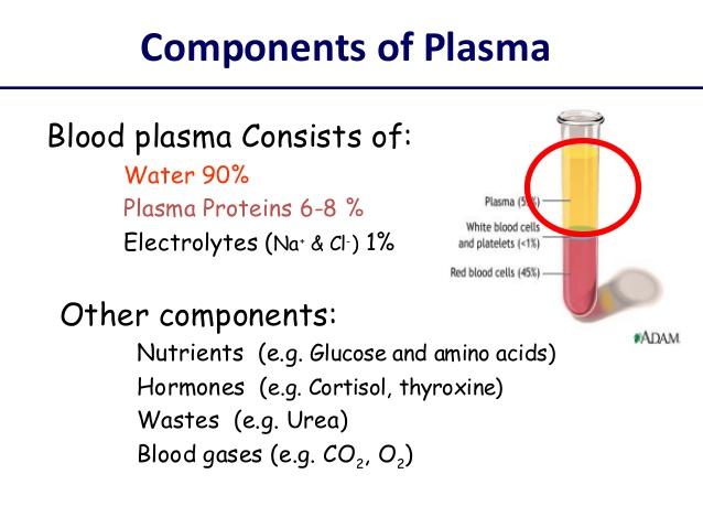
J7: Components of the Plasma.
Plasma is a pale yellow sticky liquid. It makes up 55% of the blood's volume. Thecomponents of plasma are water 92%, dissolved protein 8%, glucose, amino acids, vitamins, minerals, urea, uric acid, CO2, hormones, antibodies.
https://www.youtube.com/watch?v=BEWjOCVEN7M
J7: Components of the Plasma.
Plasma is a pale yellow sticky liquid. It makes up 55% of the blood's volume. Thecomponents of plasma are water 92%, dissolved protein 8%, glucose, amino acids, vitamins, minerals, urea, uric acid, CO2, hormones, antibodies.
J8: Give functions for the following.
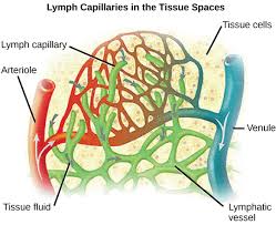
Lymph capillaries are tiny, thin-walled vessels located in the spaces between cells (except in the central nervous system and non-vascular tissues) which serve to drain and process extra-cellular fluid.
Veins are an important part of our circulatory system. They are responsible for returning deoxygenated blood back to the heart after arteries carry blood out.
Lymph nodes are major sites of B, T, and other immune cells. Lymph nodes are important for the proper functioning of the immune system, acting as filters for foreign particles and cancer cells.
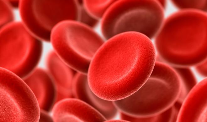
J9: Describe Red Blood Cells
Red blood cells (RBCs), also called erythrocytes, are the most common type of blood cell and the vertebrate organism's principal means of delivering oxygen (O2) to the body tissues—via blood flow through the circulatory system. RBCs take up oxygen in the lungs or gills and release it into tissues while squeezing through the body's capillaries.
Red blood cells (RBCs), also called erythrocytes, are the most common type of blood cell and the vertebrate organism's principal means of delivering oxygen (O2) to the body tissues—via blood flow through the circulatory system. RBCs take up oxygen in the lungs or gills and release it into tissues while squeezing through the body's capillaries.
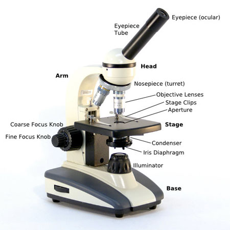
J10: Correct use of a compound microscope.
- Turn the revolving turret (2) so that the lowest power objective lens (eg. 4x) is clicked into position.
- Place the microscope slide on the stage (6) and fasten it with the stage clips.
- Look at the objective lens (3) and the stage from the side and turn the focus knob (4) so the stage moves upward. Move it up as far as it will go without letting the objective touch the coverslip.
- Look through the eyepiece (1) and move the focus knob until the image comes into focus.
- Adjust the condenser (7) and light intensity for the greatest amount of light.
- Move the microscope slide around until the sample is in the centre of the field of view (what you see).
- Use the focus knob (4) to place the sample into focus and readjust the condenser (7) and light intensity for the clearest image (with low power objectives you might need to reduce the light intensity or shut the condenser).
- When you have a clear image of your sample with the lowest power objective, you can change to the next objective lenses. You might need to readjust the sample into focus and/or readjust the condenser and light intensity. If you cannot focus on your specimen, repeat steps 3 through 5 with the higher power objective lens in place. Do not let the objective lens touch the slide!
- When finished, lower the stage, click the low power lens into position and remove the slide.
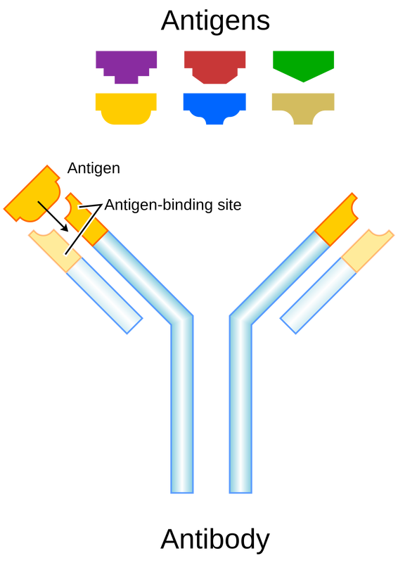
J11: Role of Antigens and Antibodies.
Antibodies are the most common and the most important. They circulate in the blood and other body fluids, defending against invading bacteria and viruses. The binding of antibodies with bacterial or viral antigens activates other immune cells that engulf and destroy the antigens.
Antibodies are the most common and the most important. They circulate in the blood and other body fluids, defending against invading bacteria and viruses. The binding of antibodies with bacterial or viral antigens activates other immune cells that engulf and destroy the antigens.
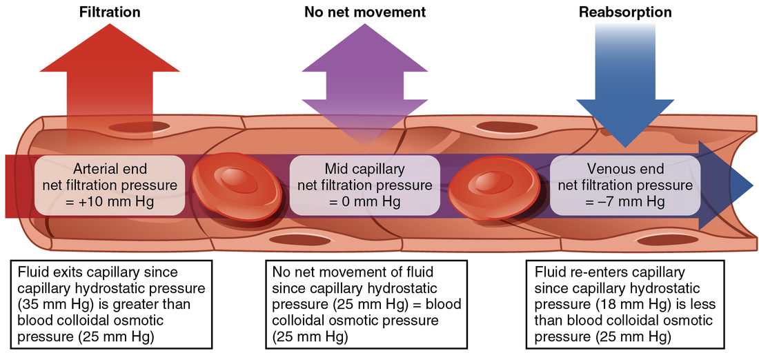
J12: Explain Capillary-tissue fluid exchange.
Where does Capillary Tissue Fluid Exchange occurIn capillary beds where blood is in close proximity - within one endothelial layer, to tissues throughout the whole body
What is the purpose of Capillary tissue fluid exchageOxygen and nutrients are able to enter tissue fluid from the blood and CO2 and metabolic wastes leave the tissue fluid and enter the blood
What is the reason for the decrease in pressure and blood velocity as it moves away from the heart. Why is this necessartthe branching of arteries into arterioles and then capillaries increases the cross sectional area of blood
Allows time for tissue fluid exchange