K1: List the following functions.
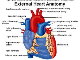
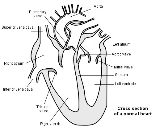
Left and Right Atria: Oxygen-rich blood from the lungs enters the left atrium through the pulmonary vein. The blood is then pumped into the left ventricle chamber of the heart through the mitral valve. From there, the blood is ready to be pumped into the body to deliver oxygen-rich blood to all bodily tissues.
Left and Right Ventricles: The right ventricle is the chamber within the heart that is responsible for pumping oxygen-depleted blood to the lungs. The right ventricle is one of the heart's four chambers. It is located in the lower right portion of the heart below the right atrium and opposite the left ventricle.
Coronary Veins: The coronary sinus is a collection of veins joined together to form a large vessel that collects blood from the heart muscle (myocardium). It delivers deoxygenated blood to the right atrium, as do the superior and inferior vena cavae.
Aorta: The aorta is the largest artery in the body. The aorta begins at the top of the left ventricle, the heart's muscular pumping chamber. The heart pumps blood from the left ventricle into the aorta through the aortic valve.
Pulmonary Trunk: The pulmonary trunk is a major vessel of the human heart that originates from the right ventricle. It branches into the right and left pulmonary arteries, which lead to the lungs.
Semilunar Valve: The semilunar valves act to prevent backflow of blood from the arteries to the ventricles during ventricular diastole, and to help maintain pressure on the major arteries. The aortic semilunar valve separates the left ventricle from the opening of the aorta.
Septum: When it is between the lower or pumping chambers (ventricles) it is called the ventricular septum. The septum keeps blood from the right and left sides of the heart from mixing. This is important because the blood in the left ventricle is loaded with oxygen for the rest of the body to use.
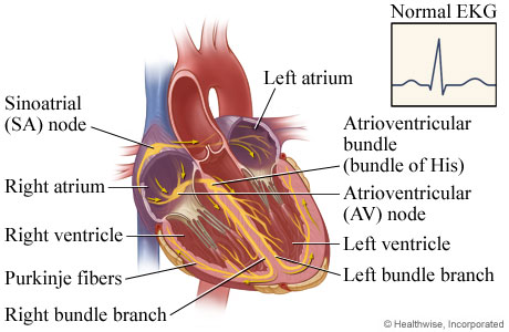
K2: Describe AV Node, SA Node, and Purkinje fibers.
AV Node: The AV Node takes the signal from the Sinoatrial (SA) Node, slows the signal down and regulates it, and then sends the electrical impulses from the atria to the ventricles (bundle of His).
SA Node: The SA node is the heart's natural pacemaker. The SA node consists of a cluster of cells that are situated in the upper part of the wall of the right atrium (the right upper chamber of the heart). The electrical impulses are generated there. The SA node is also called the sinus node.
Purkinje Fibers: Purkinje fibers are a unique cardiac end-organ. Further histologic examination reveals that these fibers are split in atria and ventricles walls. The electrical origin of atrialPurkinje fibers arrives from the sinoatrial node.

K3: Describe the automatic regulation of the heartbeat by the nervous system.
The autonomic system has two divisions: the parasympathetic system, and the sympathetic system. The parasympathetic system causes the heartbeat to slow down, and the sympathetic system causes the heartbeat to speed up. Factors such as the relative need for oxygen or blood pressure determine which of these systems is activated.
K4: Relate factors that affect and regulate blood pressure to hypertension and hypotension.
Hypertension is systolic pressure, the highest arterial pressure.
Hypotension is diastolic pressure, the lowest arterial pressure.
-large cross-sectional area
-large distance from left ventricle
K5 and K6: Demonstrate the measurement of blood pressure.
The doctor measures the maximum pressure (systolic) and the lowest pressure (diastolic) made by the beating of the heart.
K7: Diagram of Heart internally and the outside
The doctor measures the maximum pressure (systolic) and the lowest pressure (diastolic) made by the beating of the heart.
- The systolic pressure is the maximum pressure in an artery at the moment when the heart is beating and pumping blood through the body.
- The diastolic pressure is the lowest pressure in an artery in the moments between beats when the heart is resting.
K7: Diagram of Heart internally and the outside